14 Major Sulci
Main sulci are formed early in development
Fissures = really deep sulci
Typically Continuous Sulci
- Interhemispheric fissure
- Sylvian fissure
- Parietal-occipital fissure
- Calcarine sulcus
- Collateral sulcus
- Central sulcus
Typically Discontinuous Sulci
- Cingulate sulcus
- Precentral sulcus
- Postcentral sulcus
- Intraparietal sulcus
- Superior temporal sulcus
- Inferior temporal sulcus
- Superior frontal sulcus
- Inferior frontal sulcus
Other minor sulci are much less reliable
(Image from Ono, Atlas of the Cerebral Sulci, p.7)
Interhemispheric Fissure
- Hugely deep
- Divides brain into 2 hemispheres
Sylvian Fissure
- Hugely deep
- Mostly horizontal
- Insula (purple) is buried within it
- Separates temporal lobe from parietal and frontal lobe
Parietal-occipital Fissure and Calcarine Sulcus
Parietal-occipital Fissure (red):
- Very deep
- Often Y-shaped from sagittal view
- X-shaped in horizontal and coronal views
Cuneus (pink):
- Visual areas on medial side above
- Calcarine (lower visual field)
Calcarine Sulcus (blue):
- Contains V1
Lingual gyrus (yellow):
- Visual areas on medial side below calcarine and above collateral sulcus (upper visual field)
Collateral Sulcus
- Divides lingual (yellow) and parahippocampal (green) gyri from fusiform gyrus (pink)
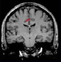
Cingulate Sulcus
- Divides the cingulate gyrus (turquoise) from precuneus (purple) and paracentral lobule (gold)
Central, Postcentral, and Precentral Sulci
Central Sulcus (red):
- Usually freestanding (no intersections)
- Just anterior to ascending cingulate
Precentral Sulcus (green):
- Often in two parts (superior and inferior)
- Intersects with superior frontal sulcus (T-junction)
- Marks the anterior end of precentral gyrus (motor strip, yellow)
Postcentral Sulcus (blue):
- Often in two parts (superior and anterior)
- Often intersects with intraparietal sulcus
- Marks posterior end of postcentral gyrus (somatosensory strip, purple)
Intraparietal Sulcus
- Anterior end usually intersects with inferior postcentral (some texts call inferior postcentral the ascending intraparietal sulcus)
- Posterior end usually forms a T-junction with the transverse occipital sulcus (just posterior to the parieto-occipital fissure)
- IPS divides the superior parietal lobule from the inferior parietal lobule (angular gyrus, gold, and supramarginal gyrus, lime)
Slice Views
- Central sulcus = red
- Precentral sulcus = green
- Transverse-occipital = purple
- Postcentral sulcus = blue
- Intraparietal sulcus = yellow
Superior and Inferior Temporal Sulci
Superior Temporal Sulcus (red)
- Divides the superior temporal gyrus (peach) from middle temporal gyrus (lime)
Inferior Temporal Sulcus (blue)
- Not usually very continuous
- Divides middle temporal gyrus from inferior temporal gyrus (lavender)
Superior and Inferior Frontal Sulci
Superior Frontal Sulcus (red)
- Divides superior frontal gyrus (mocha) from the middle frontal gyrus (pink)
Inferior Frontal Sulcus (blue)
- Divides the middle frontal gyrus from the inferior frontal gyrus (gold)
**Orbital gyrus (green) and frontal pole (grey) also are shown.
Medial Frontal View
- Superior frontal gyrus continues on medial side
- Frontal pole (grey) and frontal orbital gyrus (green) also shown


